Scientists from the Department of Optics at the Palacký University Faculty of Science have introduced a new microscopic technique, marking a fundamental shift in the rules of cellular imaging. It allows for rapid, contactless, and non-invasive analysis of cells in high definition. Their results have been published in the prestigious scientific journal Optica.
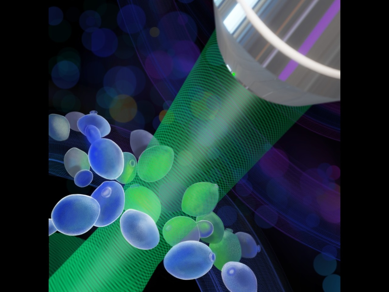
Their method allows capturing the interior structure of cells with a resolution of 250 nanometres and a phase accuracy below 2 nanometres. It can thus capture optical path differences corresponding to the size of just a few molecules. In addition, it works with conventional non-coherent illumination and thus eliminates artifact interference, allowing for sharper and more rapid imaging. Thanks to the new optical arrangement, the method is vibration resistant and ideal for consistent time-lapse measurements.
“We have shown that lateral shearing digital holographic microscopy can work even in conventional lighting, with shearing distances exceeding the coherence area of the illumination source. By doing so, we are opening up new possibilities for rapid, contactless, and non-invasive analysis of cells with high precision,” said the article’s main author, Jaromír Běhal.
The successful publication of their research results is a springboard for the resolution of a Postdoc Individual Fellowship Incoming grant by the Grant Agency of the Czech Republic – MAGICY (Machine‑learning Assisted tomoGraphic phase Imaging flow CYtometry) – which Jaromír Běhal received after returning from a two-and-a-half-year postdoctoral stay in the group under Prof Pietro Ferraro in Naples.
The new research group, Advanced Functional Imaging (AFI), under Běhal’s helm will now focus on connecting quantitative phase microscopy with flow tomographic cytometry and AI. Their goal is to detect and sort individual cells in real time and create their 3D models within a fraction of a second.
“The newly-developed technology makes possible cellular analysis without the use of staining agents to obtain precise, nanometre-accurate information on the thickness and refractive index of subcellular structures,” said Miroslav Ježek, head of the QOLO group. “Jaromír has done a tremendous job. In just a short time after returning from Italy, he has built his own team and been awarded an important grant. For our QOLO group, it is a great honour to be there with him and to support his start as a junior group leader,” Ježek added, in appreciation of the scientific work of his younger colleague.
Financing: The research was supported by the Czech Science Foundation (project no. 25-17712I).
Links:
QOLO Laboratory at the UP Faculty of Science Department of Optics